The Skeletal System
The skeletal system consists of bones, cartilages, and joints that form the framework of the human body. This structure provides support, protection, and enables movement. The skeleton is essential for maintaining the shape and structure of the body.
Components of the Human Skeleton
- Bones: Tough and rigid connective tissue responsible for strength and support in the human skeleton.
- Cartilage: A flexible connective tissue, softer than bone, which helps reduce friction and absorb shock in joints.
- Joints: Structures where bones, or bones and cartilages, meet, allowing movement and flexibility in the skeleton.
Functions of the Skeletal System
- Facilitates movement through limb action.
- Protects delicate organs like the brain, lungs, and heart.
- Provides mechanical support to body structures.
- Gives shape and form to the body.
- Produces red blood cells in bone marrow.
- Serves as attachment points for muscles.
- Stores minerals such as calcium and phosphorus.
Forms of Skeletons
Skeletons in animals are made of different materials and can take various forms:
- Chitin: A tough, lightweight material forming the exoskeleton of arthropods, such as insects. It provides protection but must be shed for growth through a process called molting.
- Cartilage: Found in vertebrates, cartilage is flexible and tough, aiding in support and reducing joint friction. It is found in cartilaginous fishes, human embryos, and certain adult structures like the ear and nose.
- Bones: Rigid structures that form the framework of higher animals. They consist of compact and spongy tissues and contain cells called osteocytes, which maintain bone health.
Types of Skeletons
- Hydrostatic Skeleton: Found in soft-bodied animals like worms, this skeleton relies on fluid pressure within the body to maintain shape and movement.
- Exoskeleton: A hard external skeleton, found in arthropods, made of chitin. It provides protection and must be shed during growth.
- Endoskeleton: An internal skeleton made of bones and cartilage, found in vertebrates. It provides support, protection, and facilitates movement.
Types of Cartilage
Cartilages are of three main types in mammals. They are:
- Hyaline cartilage: This is the most common type of cartilage and contains a dense meshwork. It can be found on the surface of movable joints, the trachea and bronchi (for ease of respiration), and in the protruding parts of the nose.
- Fibro-cartilage: Tougher than hyaline cartilage, it is found in the intervertebral discs of the vertebral column. Its main function is to provide support and strength to structures.
- Elastic cartilage: Found in the external ear (pinna) and epiglottis (a cartilaginous flap covering the trachea active during food swallowing), this type of cartilage is more flexible than the others.
Types of Bones
Long Bones
Long bones are essential for movement and supporting body weight against gravity. They grow significantly during development, with growth occurring at the epiphyseal plates. In children, red bone marrow in long bones produces blood cells. However, in adults, red marrow is largely replaced by yellow marrow, with blood cell production shifting to flat bones.
Examples of long bones include:
- Lower limbs: Tibia, fibula, femur, metatarsals, and phalanges
- Upper limbs: Humerus, radius, ulna, metacarpals, and phalanges
- Clavicle (collarbone)
Short Bones
Short bones are cube-shaped and provide support, strength, and mobility in complex structures like the wrists and ankles. They are held together by strong ligaments, offering stability while allowing flexibility and movement.
Examples of short bones include:
- Wrist (carpals): Scaphoid, lunate, triquetral, hamate, pisiform, capitate, trapezoid, trapezium
- Ankle (tarsals): Calcaneus, talus, navicular, cuboid, and cuneiform bones
Flat Bones
Flat bones primarily protect internal organs and do not require significant motion or growth. In adults, they are the main sites of blood cell production within red bone marrow.
Examples of flat bones include:
- Skull: Occipital, parietal, frontal, nasal, lacrimal, vomer
- Sternum (breastbone) and ribs
- Scapulae (shoulder blades)
Irregular Bones
Irregular bones have complex shapes tailored to specific functions, such as protecting organs and providing structural support.
Examples of irregular bones include:
- Vertebrae: Protect the spinal cord while allowing flexibility
- Pelvic girdle: Shields internal organs and provides a base for the legs
Vertebrate Skeleton
The vertebrate skeleton consists of two major parts:
- Axial Skeleton: Includes the skull, spinal column (vertebrae), and ribs.
- Appendicular Skeleton: Comprises the limbs (arms and legs), shoulder girdle, and pelvic girdle.
The Axial Skeleton
1. The Skull
The skull is a hollow structure made of flat bones that protect the brain and house sensory organs like the eyes and ears. It includes the jaws, which hold the teeth.
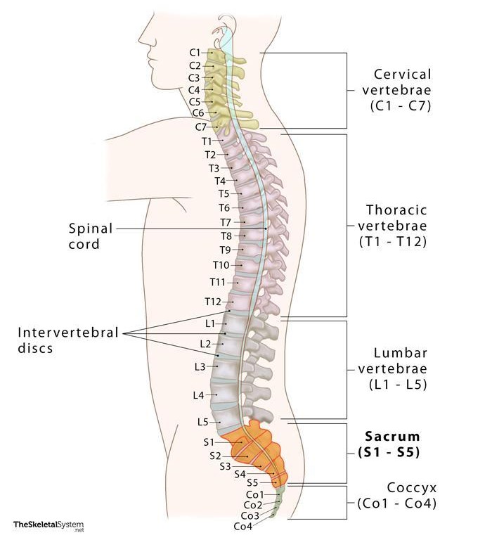
2. The Vertebral Column
The vertebral column supports the body, protects the spinal cord, and provides attachment points for ribs and muscles. It consists of 33 vertebrae divided into five regions:
- Cervical: Found in the neck, includes the atlas and axis, which allow head movement.
- Thoracic: Located in the chest, supports the ribs and aids in breathing.
- Lumbar: Found in the lower back, supports the body’s weight.
- Sacral: Located in the pelvis, provides strength and stability.
- Coccygeal: The tailbone, a fused structure providing minor support and muscle attachment.
Vertebral column
It is a flexible column of vertebrae, connecting the trunk of human body to the skull and appendages. The vertebrate are held together with strong ligament and comprehensible cartilage pads called into intervertibratal disc.
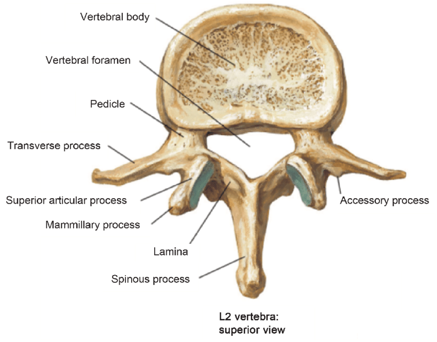
Structure of a typical Vertebrae
All the vertebrae basically have the following features in common:
- Centrum which is a solid piece of bone below the neural canal.
- Neural Arch which is carried by the Centrum
- A neural canal surrounded by both the Centrum and the arch
- Neural spine which projects upwards dorsally
- Facets for the articulation with other parts of the skeleton.
- 2 transverse processes which are sideways projections from each vertebra
- Transverse foramen
It is composed of 33 vertebrae which are divided into 5 regions: Cervical, Thoracic, Lumbar, Sacral, and Coccygeal.
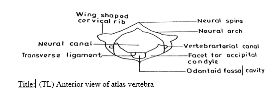
Cervical Vertebrae
The first cervical vertebra is called the atlas while the
second is called the axis.
The Atlas It has a large neural canal, flat and broad
transverse processes, short neural spine which could
be absent at times. It also has a vertebrarterial canal
for
the passage of blood vessels. Centrum is absent.
The top two vertebrae in the spinal column are
specialized
to allow the head a greater range of
movement than would be possible with normal vertebrae. A
stable ball-and-socket joint
accommodates both side-to-side and up-and-down
motion.
Lumbar Vertebrae
These are found in the upper abdominal region. In man there are 5 of them. Each has large and flat transverse processes, broad and flat neural spine, large and thick centrums and well developed zygapophyses. They provides attachment to abdominal muscles
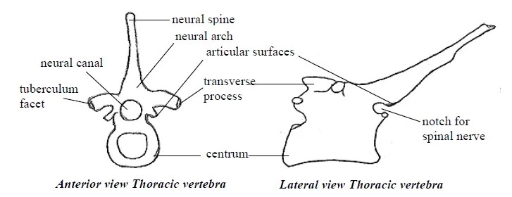
Thoracic Vertebra
These are found in the chest region. In man there are 12 of them. It has long and prominent neural spine which projects upwards and backwards, a pair of short transverse processes, a large neural canal and neural arc and large cylindrical centrums. They also have particular surfaces for attachment of the ribs. They aid in the attachment of ribs and assist in breathing alongside the ribs.
Sacral Vertebrae
This is found in the lower abdominal region. In man, they are 5 in number and fuse together to form a sacrum. Each sacral vertebrate has a narrow neural canal, reduced neural spine and large centrums.They Join the pelvic girdle to provide support,rigidity and strength.
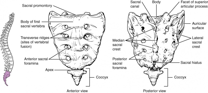
Caudal vertebrae
These are joined together to form a singular bony mass called coccyx. Each has no neural spine, no neural canal and no transverse process. they are the bones of the tail. They are 4 small fused bones which ends the vertebral column. The coccyx supports the tail and provides attachment for tail muscles
3. Rib Cage
The rib cage protects vital organs like the heart and lungs. It includes:
- True Ribs: The first seven ribs directly attached to the sternum.
- False Ribs: The next three ribs connected to the sternum through cartilage.
- Floating Ribs: The last two pairs of ribs, which are not attached to the sternum.
The Appendicular Skeleton
1. Pectoral Girdle
The pectoral girdle connects the arms to the axial skeleton. It includes the scapulae (shoulder blades) and clavicles (collarbones).
2. Pelvic Girdle
The pelvic girdle supports the lower body and transfers the body’s weight to the legs. It consists of two pelvis bones connected to the sacrum and joined at the front by fibro-cartilage.
3. Limbs
Both the forelimbs (arms) and hind limbs (legs) follow a pentadactyl plan. Each limb consists of:
- A long bone (e.g., humerus in arms, femur in legs).
- Two parallel long bones (e.g., radius and ulna in arms, tibia and fibula in legs).
- Small bones (e.g., carpals in wrists, tarsals in ankles).
- Digit bones (phalanges) forming fingers and toes.
Joints
Joints are points where two bones meet, allowing movement. They are cushioned by cartilage and lubricated with synovial fluid to reduce friction.
Classification of Joints
Joints can be classified in two ways:
Structural Classification
This classification is based on how bones are connected. Bones may be joined by fibrous tissue, cartilage, or within a fluid-filled cavity. The three types of joints in this category are:
- Fibrous joints: Connected by fibrous tissue
- Cartilaginous joints: Connected by cartilage
- Synovial joints: Connected within a fluid-filled cavity
Types of Joints
- Immovable Joints: Found in the skull, these joints do not allow movement.
- Movable Joints: These joints allow
a range of movements, including:
- Hinged Joints: Allow movement in one plane, e.g., knees and elbows.
- Ball-and-Socket Joints: Allow multi-directional movement, e.g., shoulders and hips.
- Gliding Joints: Allow sliding movements, e.g., wrists and ankles.
synarthroses
Immovable joints, also known as synarthroses, are fixed joints that provide a strong connection between the bones they join.
These joints are primarily fibrous or cartilaginous and are located in areas where they safeguard vital organs such as the brain and heart.
Examples of immovable joints include skull sutures, gomphosis joints, and the manubriosternal joint, a cartilaginous joint linking the sternum to the manubrium, offering protection to the heart.
amphiarthroses
Slightly movable joints, also known as amphiarthroses, allow limited movement. These joints are typically found between vertebrae in the spine.
One example is the intervertebral discs, which connect the vertebrae and permit slight motion between the bones of the spine.
Another example is the pubic symphysis, located in the pelvic region.
Diarthroses
Freely movable joints, also known as diarthroses, are synovial joints that provide the greatest range of motion. They are primarily located in the limbs and enable various types of movements.
Based on the number of movement axes, synovial joints are classified as follows:
- Uniaxial joints: These allow movement in a single plane. For example, the elbow joint permits only bending and straightening.
- Biaxial joints: These allow movement in two planes. For instance, knuckle joints enable bending and straightening of the fingers, as well as spreading them apart.
- Multiaxial joints: These allow movement in multiple directions along all three axes. Examples include the shoulder and hip joints, which enable forward, backward, sideways, and rotational movements.
Synovial joints support the following movements:
- Abduction: Moving a limb away from the body's midline.
- Adduction: Moving a limb toward the body's midline.
- Extension: Straightening a limb at a joint.
- Flexion: Bending a limb at a joint.
- Rotation: Circular movement around a fixed point.
Fibrous Joints
Fibrous joints are tightly held together by a thin layer of strong connective tissue, typically collagen. These joints are fixed, meaning they allow no movement, and do not have a joint cavity. They are also known as fixed or immovable joints, primarily providing structural stability.
Examples of Fibrous Joints
-
Sutures: These consist of dense
fibrous connective tissue joining specific bones of
the skull. Prominent sutures include:
- Coronal suture
- Sagittal suture
- Lambdoid suture
- Squamous suture
- Gomphoses: Known as peg-and-socket joints, gomphoses attach the roots of the teeth to the alveolar sockets in the lower jaw (mandible) and upper jaw (maxillae).
- Syndesmoses: These are fibrous joints found between certain long bones, such as the tibia and fibula.
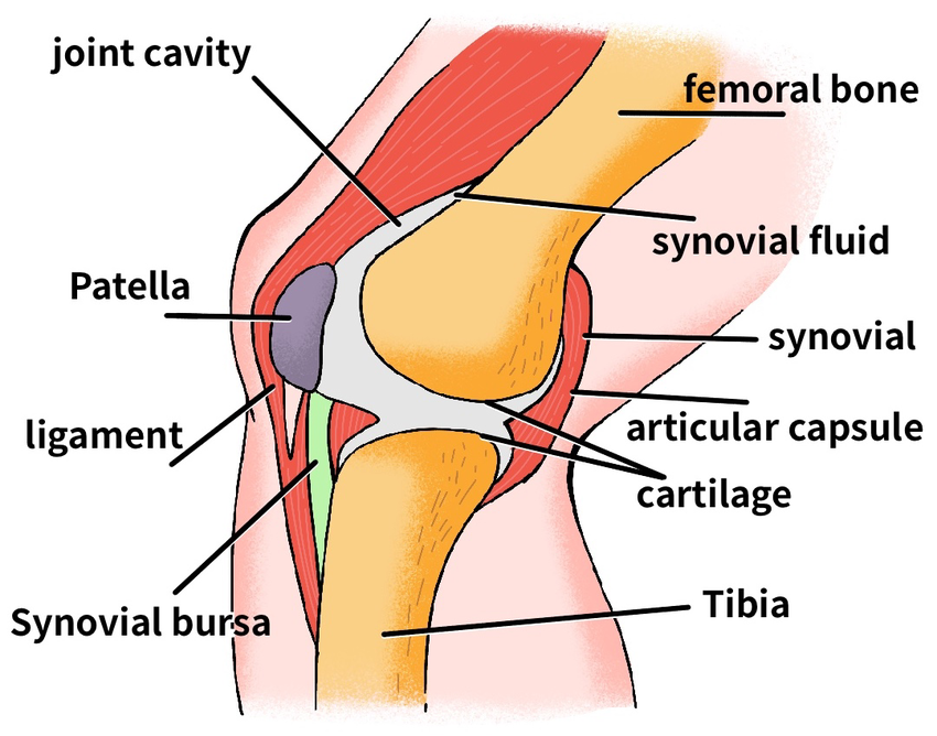
Synovial Joints
Synovial joints are the most common type of joint in the human body. They allow a wide range of motions such as walking, running, typing, and more. These joints are flexible, movable, rotatable, and capable of sliding over one another. Common examples of synovial joints include the shoulder joint, neck joint, knee joint, and wrist joint.
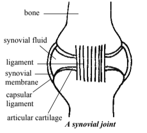
Characteristics of Synovial Joints
- The ends of the bones are covered with a layer of smooth hyaline cartilage, called articular cartilage. This reduces friction at the joint.
- The joint is enclosed by a bag-like capsular ligament, which holds the joint together and contains synovial fluid.
- The capsular ligament is lined with a synovial
membrane, which:
- Secretes synovial fluid into the synovial cavity.
- Acts as a seal to waterproof the joint.
- Lubricates the joint and reduces wear and tear caused by friction.
- Strong, tough ligaments made of dense connective tissue attach and hold the bones together. These ligaments prevent dislocation during normal movement.
- The articulating surfaces of adjacent bones are shaped to fit together reciprocally, ensuring smooth movement.